


Lipoleiomyoma of the ovary – a case report and a literature review
Dobrosława L. Sikora-Szczęśniak1, Grzegorz Szczęśniak2
 Affiliacja i adres do korespondencji
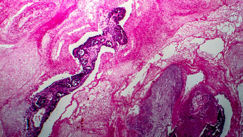
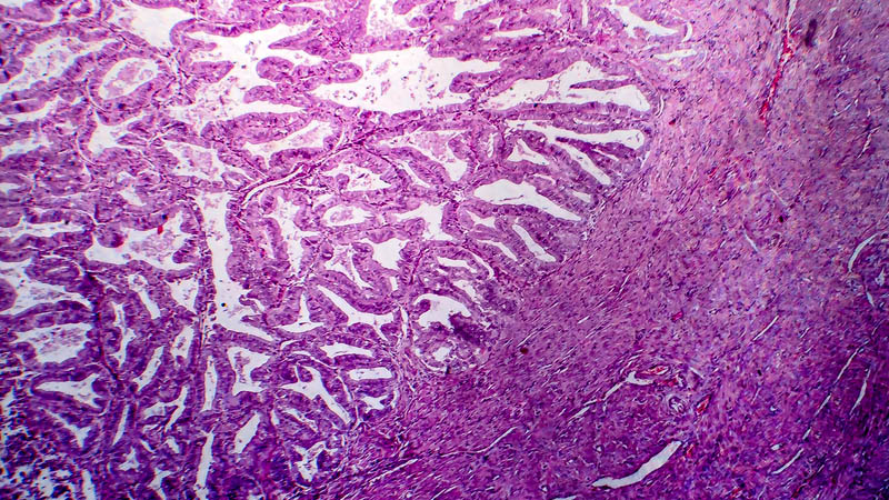
Leiomyoma belongs to rare benign ovarian tumors. Cellular leiomyoma (8892/0) and lipoleiomyoma (8890/0) are histological variants of leiomyoma (8890/3), which is the most common benign tumor in women. Cellular leiomyoma is found in about 5% of uterine leiomyoma cases. Histologically, it presents as a highly cellular tissue with increased cellular density compared to myometrium. The cells are usually small, spindle-shaped, without atypia, with low mitotic activity (<5 mitotic figures per 10 HPF) and scarce connective tissue component. Lipoleiomyoma is composed of adipocytes, smooth muscle cells and fibrous tissue. Only single cases of ovarian lipoleiomyoma are documented in the available English-language literature. We present a case of a 51-year-old patient qualified for laparotomy due to right ovarian tumor. A tumor about 70 mm in diameter was excised followed by hysterectomy with bilateral salpingo-oophorectomy. Postoperative histopathological evaluation revealed right ovarian tumor (lipoleiomyoma cellulare oedematosum); in the uterine corpus – leiomyomata intramuralia partim cellularia et leiomyoma submucosum corporis uteri. So far, there have been no case reports of cellular lipoleiomyoma of the ovary (lipoleiomyoma cellulare) in the literature. Patients after surgical procedures due to rare histological subtypes of ovarian leiomyoma should remain under long-term clinical surveillance.