


SestaMIBI scintigraphy in diagnosis of ovarian cancer’s relapse
Agnieszka Timorek-Lemieszczuk1, Ewa Kurowska-Mroczek1, Beata Osuch1, Maciej Jakuciński2
 Affiliacja i adres do korespondencji
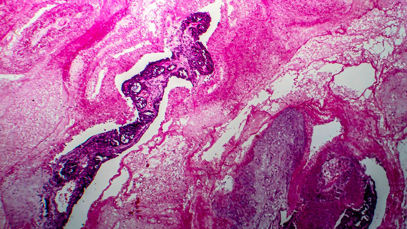
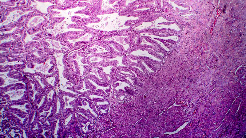
Ovarian cancer is the main cause of mortality among women suffering from malignant genital system cancer in Poland. Early detection of possible cancer recurrence after combined first-line therapy is necessary to launch adequate additional treatment with the best result for patient. Elevated concentration of CA-125 may precede clinical symptoms of reoccurrence even by a few months. A fusion of a functional study such as the positron emission tomography with a morphological study of computer tomography (PET-CT) allows for obtaining precise data about metabolic activity as well as anatomical evaluation of how advanced the cancer process is. Aim of the study: To compare the value of scintigraphy in which Tc-99m sestaMIBI is used and the value of PET-CT in early detection of cancer recurrence among women diagnosed with ovarian cancer. Material and methods: Fourteen patients diagnosed with ovarian cancer in clinical stages from IC to IIIC underwent scintigraphy (using Tc-99m sestaMIBI) of pelvis minor and abdomen as well as PET-CT of the whole body. Diagnostic value of the Tc-99m sestaMIBI scintigraphy and PET-CT was evaluated on the basis of sensitivity, specificity, and positive and negative predictive value of these tests. Results: Functional studies such as the Tc-99m sestaMIBI scintigraphy and the positron emission tomography linked with computer tomography, can be especially useful in a oncological gynecology in cases that cannot be equivocally decided using other available diagnostic methods. However, our results will require further investigation in which a greater number of patients in a study group should be used.