


TGF-b1 cytokine and TGF-b1 receptors I, II, III expression in vulvar carcinoma
Anita Olejek1, Agnieszka Michalska1, Urszula Mazurek2, Dariusz Kuśmierz2, Tadeusz Wilczok2
 Affiliacja i adres do korespondencji
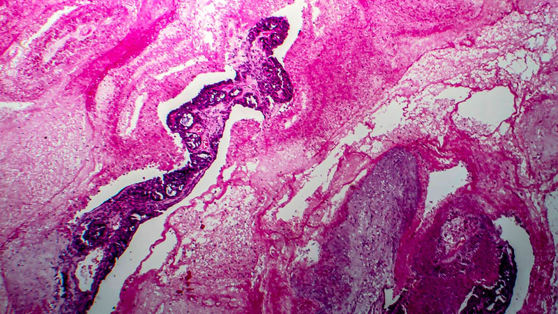
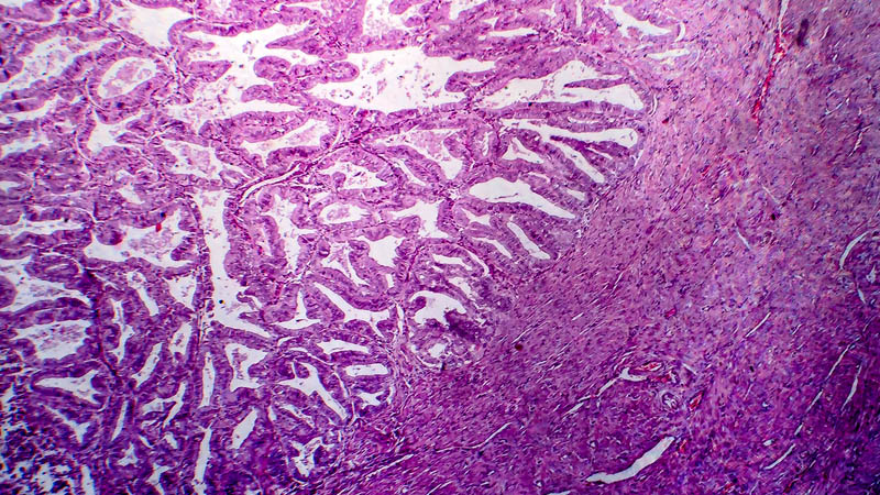
The aim of the study: Evaluation of the expression of TGF-b1 and its receptors in vulvar tissues with neoplastic lesions. Material and methods: 28 vulvar tissue samples with neoplastic lesions (16 cases of non-keratinous vulvar carcinoma, 12 – keratinous) obtained during diagnosis or surgery from patients hospitalized in Department of Obstetrics and Gynecology in Bytom. Vulvar tissue samples obtained during plastic surgery and distinguished as normal on histopathological examination were used as a control group. Histopathological examination and molecular analysis were performed on all of the samples to assess the presence of TGF-b1 and its receptors I, II, III genes. Quantitative analysis was performed using QRT-PCR (TaqMan) technique. Results: Mean number of mRNA copies in non-keratinous carcinomas was: for TGF-b1 – 9631.6, for TGFR1 – 12431.1, for TGFR2 – 12034.8, for TGFR3 – 16793.7; in keratinous carcinomas: for TGF-b1 – 14054.8, for TGFR1 – 14772.9, for TGFR2 – 8239.4, for TGFR3 – 17751.5 – in 1 mg of total RNA. The mean number of mRNA copies in 1 mg of total RNA in the control group was: TGF-b1 – 463.9, TGFR1 – 1185.9, TGFR2 – 3450.9, TGFR3 – 340.9. Conclusions: 1. The highest expression of TGFR3 gene is found in vulvar carcinoma, and in healthy tissues the highest expression was found for TGFR2 gene responsible for cell proliferation inhibition. 2. Increased expression of TGFR3 gene in planoepithelial vulvar carcinoma may indicate its influence on changes of cell growth cycle supporting carcinogenesis.